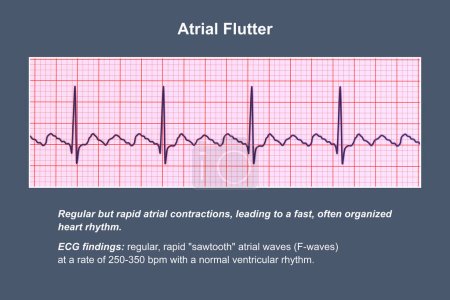
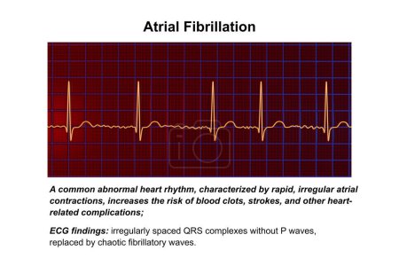
56 Bilder zum Thema "abnormal heart rhythm" bei ClipDealer

Medien sortieren nach     Gehe zu Seite:    
« Vorherige  1  Nächste »
A 3D scientific illustration depicting an electrocardiogram ECG displaying the chaotic rhythm of ventricular fibrillation, a life-threatening cardiac arrhythmia.
ECG displaying the chaotic rhythm of ventricular fibrillation, a life-threatening cardiac arrhythmia, 3D illustration.
A detailed 3D illustration of an electrocardiogram displaying sinus bradycardia, a condition characterized by a slow heart rate originating from the sinus node, typically below 60 beats per minute.
Collection of normal and abnormal ECGs. Vector of an electrocardiogram, ecg, or medical icon. A heartbeat graph
ICD Acronym or abbreviation to implantable cardioverter defibrillator as device that monitors heart rhythm problems. Word ICD is around ECG paper strips, result of echocardiography and stethoscope
Troubled senior man trying concentrating
ECG displaying the chaotic rhythm of ventricular fibrillation, a life-threatening cardiac arrhythmia, 3D illustration.
Ventricular tachycardia: A rapid heart rhythm originating in the ventricles, causes palpitations, dizziness, and life-threatening symptoms. ECG shows wide QRS complexes, 3D illustration.
ECG in atrial fibrillation (AFib), a 3D illustration depicting irregular rhythm, absent P waves, and rapid, chaotic atrial activity, posing a risk of palpitations and stroke.
A detailed 3D illustration of an Electrocardiogram ECG displaying sinus tachycardia, a regular cardiac rhythm with heart rate that is higher than the upper limit of normal of 90-100 bpm in adults.
ECG in atrial fibrillation (AFib), a 3D illustration depicts irregular rhythm, absent P waves, and rapid, chaotic atrial activity, posing a risk of palpitations and stroke.
Electrocardiogram (ECG) displaying sinus tachycardia, a regular cardiac rhythm with heart rate that is higher than the upper limit of normal of 90-100 bpm in adults, 3D illustration.
An electrocardiogram displaying sinus bradycardia, a condition characterized by a slow heart rate originating from the sinus node, typically below 60 beats per minute, 3D illustration.
ECG displaying Torsades de pointes rhythm, dangerous heart rhythm with fast, irregular beats twisting around the electrical axis, potentially causing fainting or cardiac arrest, 3D illustration
A detailed 3D illustration of an Electrocardiogram ECG displaying sinus arrhythmia, a condition characterized by irregular heart rhythms originating from the sinus node.
ECG in atrial flutter, an abnormal heart rhythm characterized by rapid, regular contractions of the atria. 3D illustration displaying characteristic sawtooth P-waves and irregular ventricular rhythm.
Electrocardiograph Traces - Cardiac Arrhythmia
Garland with red hearts lie on electrocardiogram with stethoscope head, as if listening on one of the cut out hearts. Idea for interpretation of diseases associated with impaired heart rate or pulse
Figure heart is among roasted coffee beans. Effects of coffee and caffeine on heart function, heartbeat, rhythm, and role in development of heartburn, attack, arrhythmia, failure and other diseases
Normal stage AV block in coronary diseas
Black doctor stethoscope and stress ball in heart shape on woode
All degree AV block in coronary disease
Second degree AV block in coronary disea
Third degree AV block in coronary diseas
Arrhythmia. Medical Concept on Green Background.
The abnormal fast human heart increased heart rhythm rate at which it was in cardiac arrest, anxiety, or panic attacks, 3D Render
heart and cardiac pacemaker on white background 3d render, illustration heart anatomy, section, right and left ventricle, atria, valves
Myocarditis text on textured grunge copper and vintage gold background
Arrhythmia - Inscription in Anamnesis. 3D Render.
Arrhythmias - Wording in Anamnesis. 3D Render.
Arrhythmia - Phrase in Differential Diagnoses. 3D Render.
Arrhythmia. Medicine. 3D Illustration.
3D illustration of ICD International Classification of Diseases title on a medical document
ECG in atrial fibrillation (AFib), a 3D illustration depicting irregular rhythm, absent P waves, and rapid, chaotic atrial activity, posing a risk of palpitations and stroke.
Tired asian woman suffering Thyrotoxicosis,overactive thyroid,fast heartbeat,patient with fingertip pulse oximeter checking heart rate,anxious and panic attacks due to Hyperthyroidism,health care
ECG ElectroCardioGraph paper that shows inferolateral T wave changes for clinical correlation, Resting Heart Rate HR 65 beats per minute, Abnormal ECG for a 41 years old male, Electrocardiogram
Early afterdepolarization is an arrhythmogenic mechanism that triggers activity, occurring before the end of the T wave and commonly seen in QT interval prolongation.
ECG ElectroCardioGraph paper that shows inferolateral T wave changes for clinical correlation, Resting Heart Rate HR 65 beats per minute, Abnormal ECG for a 41 years old male, Electrocardiogram
ECG in atrial fibrillation (AFib), a 3D illustration depicts irregular rhythm, absent P waves, and rapid, chaotic atrial activity, posing a risk of palpitations and stroke.
ECG ElectroCardioGraph paper that shows inferolateral T wave changes for clinical correlation, Resting Heart Rate HR 65 beats per minute, Abnormal ECG for a 41 years old male, Electrocardiogram
A detailed 3D illustration of an Electrocardiogram ECG displaying sinus arrhythmia, a condition characterized by irregular heart rhythms originating from the sinus node.
ECG ElectroCardioGraph paper that shows inferolateral T wave changes for clinical correlation, Resting Heart Rate HR 65 beats per minute, Abnormal ECG for a 41 years old male, Electrocardiogram
ECG in atrial flutter, an abnormal heart rhythm characterized by rapid, regular contractions of the atria. 3D illustration displaying characteristic sawtooth P-waves and irregular ventricular rhythm.
Abnormal ECG refers to changes in depolarization waves and or repolarization waves, most of which are pathologic and few are physiological.
ECG ElectroCardioGraph paper that shows inferolateral T wave changes for clinical correlation, Resting Heart Rate HR 65 beats per minute, Abnormal ECG for a 41 years old male, Electrocardiogram
A detailed 3D illustration of an electrocardiogram displaying sinus bradycardia, a condition characterized by a slow heart rate originating from the sinus node, typically below 60 beats per minute.
ECG ElectroCardioGraph paper that shows sinus rhythm abnormality of right ventricular hypertrophy, inferior T wave due to hypertrophy and ischemia, Abnormal ECG study, unconfirmed diagnosis
Abnormal ECG refers to changes in depolarization waves and or repolarization waves, most of which are pathologic and few are physiological.
Female, 5-year-old, clinically diagnosed with dextrocardia. The characteristic of dextrocardia electrocardiogram is a gradual decrease in R-wave amplitude from leads V1 to V6.
Electrocardiogram displaying a junctional rhythm, which occurs when the electrical signals in the heart originate from the atrioventricular junction instead of the sinoatrial node, 3D illustration.

« Vorherige  1  Nächste »