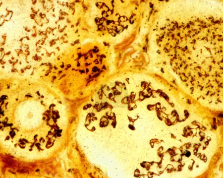
High magnification micrograph of pseudounipola ... 

Media-ID: B:720475068
Nutzungsrecht:
Kommerzielle und redaktionelle Nutzung
High magnification micrograph of pseudounipolar neurons of a dorsal root ganglion stained with the Cajal's formol-uranium silver method that demonstrates the Golgi apparatus. It appears as a brown network located in the neuron cell body around the nu
| Vorschau |
Varianten
Mediainfos
|
Dieses Bild mit unserem Kundenkonto ab €0.95 herunterladen!
|
||||
| Standardlizenz: JPG | ||||
| Format | Bildgröße | Downloads | ||
|
Print XXL 15 MP |
3840x3072 Pixel 32.51x26.01 cm (300 dpi) |
1 | ||
| Standardlizenz: JPG | ||||
| Format | Bildgröße | Netto | Brutto | Preis |
|
Web S 0.5 MP |
500x400 Pixel 16.93x13.55 cm (75 dpi) |
€3.90 | €4.17 | |
|
Print M 2 MP |
1000x800 Pixel 8.47x6.77 cm (300 dpi) |
€6.90 | €7.38 | |
|
Print XL 8 MP |
2000x1600 Pixel 16.93x13.55 cm (300 dpi) |
€12.90 | €13.80 | |
|
Print XXL 15 MP |
3840x3072 Pixel 32.51x26.01 cm (300 dpi) |
€15.90 | €17.01 | |
| Merchandisinglizenz: JPG | ||||
| Format | Bildgröße | Netto | Brutto | Preis |
|
Print XXL 15 MP |
3840x3072 Pixel 32.51x26.01 cm (300 dpi) |
€79.90 | €85.49 | |
| Media-ID: | B:720475068 |
| Aufrufe: | 1 |
| Beschreibung: | High magnification micrograph of pseudounipolar neurons of a dorsal root ganglion stained with the Cajal's formol-uranium silver method that demonstrates the Golgi apparatus. It appears as a brown network located in the neuron cell body around the nu |
Nutzungslizenz
| Nutzungsrecht: | Kommerzielle und redaktionelle Nutzung |
Userinfos
| Hinzugefügt von: | [email protected] |
| Weitere Medien von [email protected] |
Bewertung
| Bewertung: |
|
Suchbegriffe
| Keywords: |


