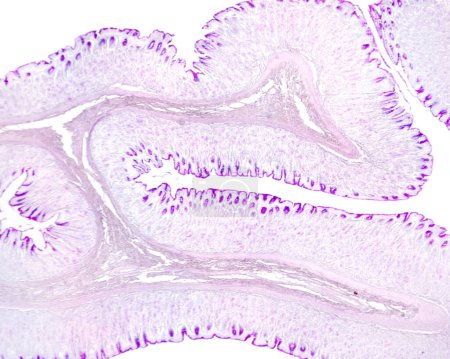
Very low magnification light micrograph of the ... 

Media-ID: B:714203744
Nutzungsrecht:
Kommerzielle und redaktionelle Nutzung
Very low magnification light micrograph of the gastric wall stained with PAS method. The inner layer is the mucosa that shows many folds. In the axis of these folds is the submucosa. In the mucosa, the mucous surface epithelium and foveolar cells of
| Vorschau |
Varianten
Mediainfos
|
Dieses Bild mit unserem Kundenkonto ab €0.95 herunterladen!
|
||||
| Standardlizenz: JPG | ||||
| Format | Bildgröße | Downloads | ||
|
Print XXL 15 MP |
3840x3072 Pixel 32.51x26.01 cm (300 dpi) |
1 | ||
| Standardlizenz: JPG | ||||
| Format | Bildgröße | Netto | Brutto | Preis |
|
Web S 0.5 MP |
500x400 Pixel 16.93x13.55 cm (75 dpi) |
€3.90 | €4.17 | |
|
Print M 2 MP |
1000x800 Pixel 8.47x6.77 cm (300 dpi) |
€6.90 | €7.38 | |
|
Print XL 8 MP |
2000x1600 Pixel 16.93x13.55 cm (300 dpi) |
€12.90 | €13.80 | |
|
Print XXL 15 MP |
3840x3072 Pixel 32.51x26.01 cm (300 dpi) |
€15.90 | €17.01 | |
| Merchandisinglizenz: JPG | ||||
| Format | Bildgröße | Netto | Brutto | Preis |
|
Print XXL 15 MP |
3840x3072 Pixel 32.51x26.01 cm (300 dpi) |
€79.90 | €85.49 | |
| Media-ID: | B:714203744 |
| Aufrufe: | 1 |
| Beschreibung: | Very low magnification light micrograph of the gastric wall stained with PAS method. The inner layer is the mucosa that shows many folds. In the axis of these folds is the submucosa. In the mucosa, the mucous surface epithelium and foveolar cells of |
Nutzungslizenz
| Nutzungsrecht: | Kommerzielle und redaktionelle Nutzung |
Userinfos
| Hinzugefügt von: | [email protected] |
| Weitere Medien von [email protected] |
Bewertung
| Bewertung: |
|
Suchbegriffe
| Keywords: |


